


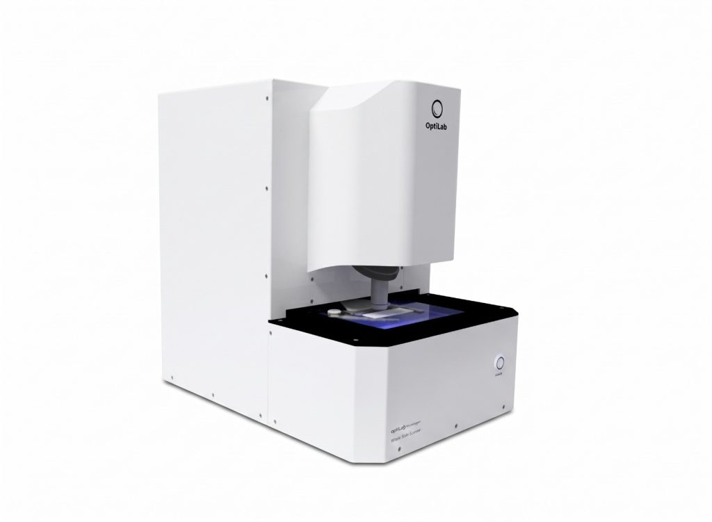
Microscopic observation is generally performed by viewing a sample through a specific field of view. However, when comprehensive documentation of the specimen area is required, taking an image from a single field of view may not be sufficient. For this purpose, technology whole slide scanner dapat menjadi pilihan.
What Is a Whole-Slide Scanner?
Whole slide scanner It is a device used to scan and document the entire image of a microscope slide. Unlike documentation methods that capture only a single field of view, this device scans the entire slide so that the entire slide can be recorded as a digital image.
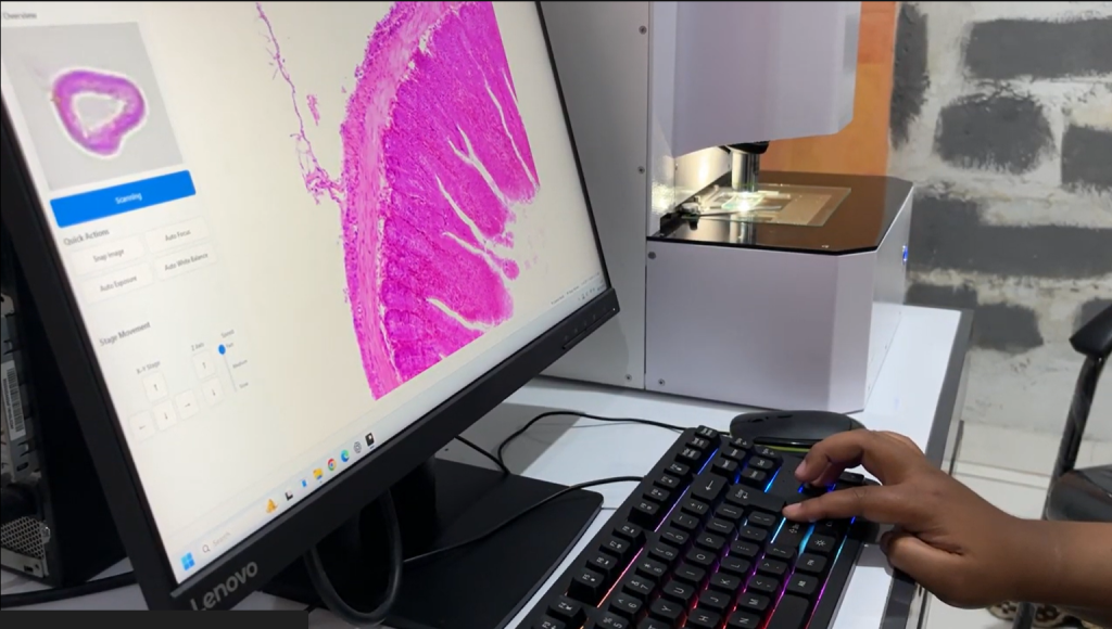
With this approach, users can obtain documentation that covers the entire specimen more comprehensively, rather than just the specific portion visible through the microscope’s field of view.
Getting to Know the OptiLab Microstepper
OptiLab Microstepper merupakan perangkat whole slide scanner from OptiLab, which is used to document images of microscope slides using a camera scanning method.
Through this scanning process, the Microstepper can capture an image of the entire specimen, so documentation is not limited to a single field of view. The product information states that the entire specimen can be recorded with the same level of detail.
How Is It Different from Single-View Documentation?
In single-field-of-view documentation, the image captured covers only the area currently visible through the microscope. To view other areas, the user must shift the observation position and take additional images.
Meanwhile, whole slide scanner It is designed to document specimens in their entirety through the scanning process. As a result, the documentation provides a more comprehensive view of the sample area in a single scan sequence.
Why Is It Important to Document All Preparations?
During microscopic observation, the area of interest is not always within a single field of view. Documenting the entire slide can help users review a broader area of the sample and save their observations in digital form.
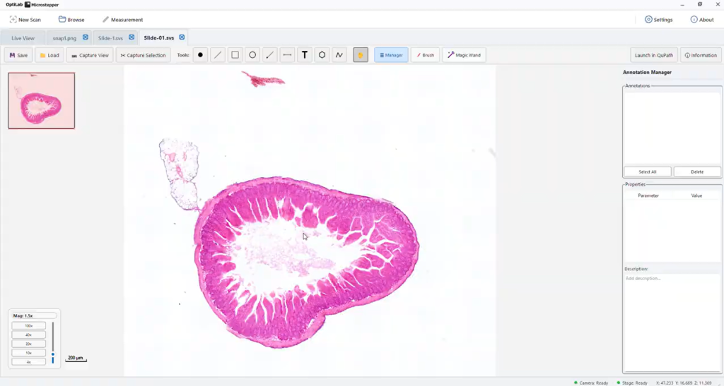
This documentation can serve as supporting material for educational and research activities, as well as for laboratory needs. Users also have image records of the specimens that can be used as references during subsequent reviews.
Supporting Microscopic Documentation with OptiLab
The need for specimen documentation continues to grow alongside the use of digital technology in laboratory activities. OptiLab Microstepper offers a solution for comprehensively scanning microscope specimens, ensuring that documentation is not limited to a single field of view.
Through this product, PT Miconos and OptiLab support the need for microscopic documentation in education, research, and laboratory settings. For product information and to make a purchase, visit PT Miconos' official website.