


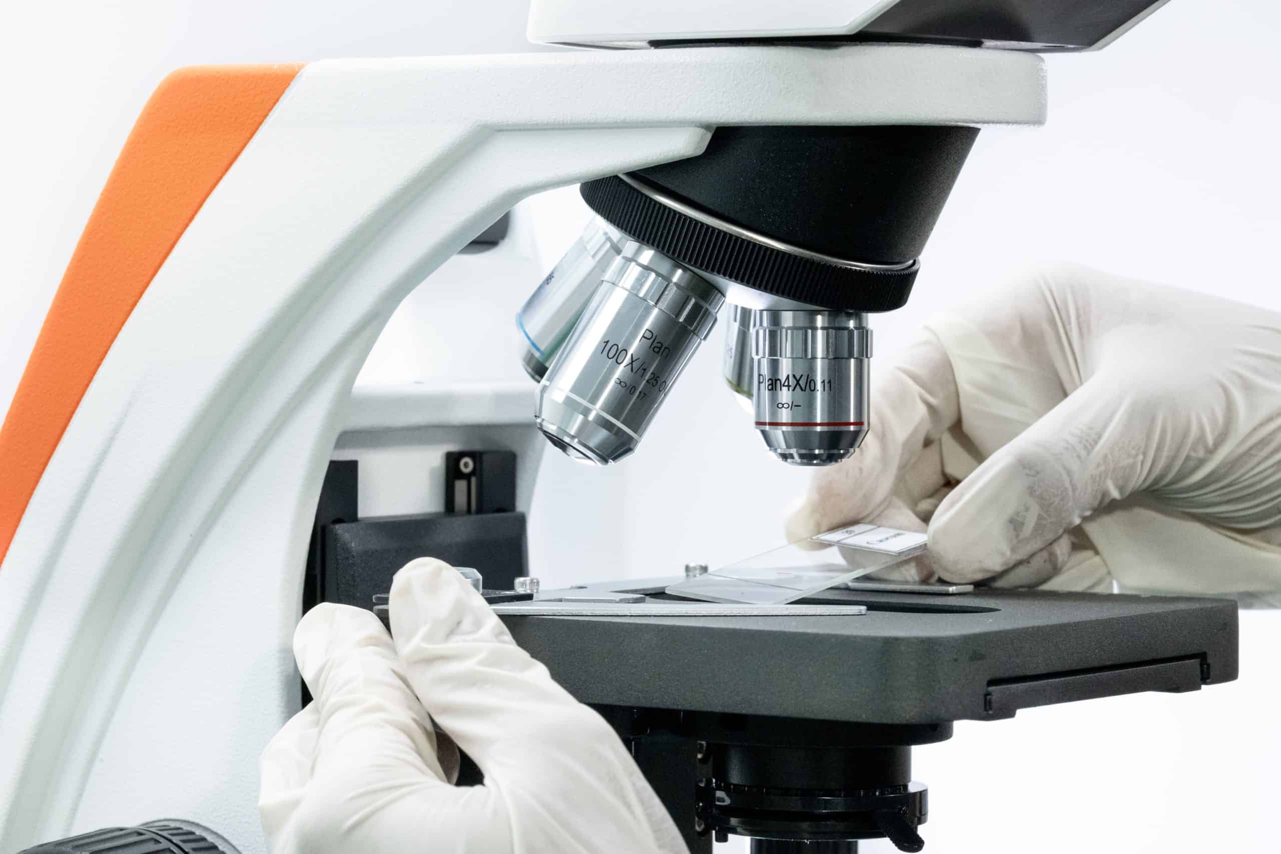
In laboratory observation activities, a microscope requires a ready-to-observe object. The object is placed on a specialized thin piece of glass so it can be illuminated and magnified by the lens. This specifically prepared object or sample is known as a microscope slide. The use of slides is crucial in biological, medical, and industrial research. Without a properly prepared specimen, the microscope lens cannot produce a focused, clear, and precise image.
A microscope slide consists of a tissue, cell, or microorganism sample placed on a glass slide(object glass). The sample is then covered with an even thinner cover slip (cover glass)
The primary purpose of slide preparation is to keep the specimen in place so it does not shift during observation. Additionally, covering the sample protects the objective lens from fluid or chemical contamination.
Types of Microscope Slides
Based on their shelf life and preparation methods, microscope slides are divided into several main categories:
Wet Mounts (Fresh Slides): The sample is suspended in a liquid such as water or a stain, then observed immediately. This type is temporary and ideal for observing the movement of living cells.
Prepared (Permanent) Slides: The sample undergoes specific fixation, dehydration, and staining processes to ensure durability. These slides are typically used for educational practicums and long-term research.
Smear Slides: A liquid sample is spread thinly across the glass slide. This method is frequently applied to blood, sputum, or bacterial sample examinations.
Sectioned Slides: Solid tissue samples are sliced very thinly using a microtome so that the microscope's light can pass through the tissue.
The Main Functions of Slides in Observation
Using the appropriate slide provides various technical advantages in laboratory analysis:
Facilitates focused observation on specific cellular or tissue structures.
Protects the specimen from damage caused by exposure to outside air during analysis.
Ensures that the illumination from the microscope condenser is distributed evenly.
Simplifies digital documentation and data analysis when connected to a microscope camera.
The working mechanism of a slide centers on light transmission. Light from the microscope's illumination source is transmitted through the glass slide, the thin sample, and the cover slip.
The refracted light from the sample is captured by the objective lens for magnification, then passed on to the eyepiece or a digital camera sensor. Because of the thin and flat sample structure, the cell image can be displayed sharply on a monitor screen or through the viewing lens.
Does your laboratory facility require updated, precision microscopes and digital cameras for research needs? Contact the PT Miconos team today to consult and get the best laboratory instrument solutions tailored to your operational targets!