


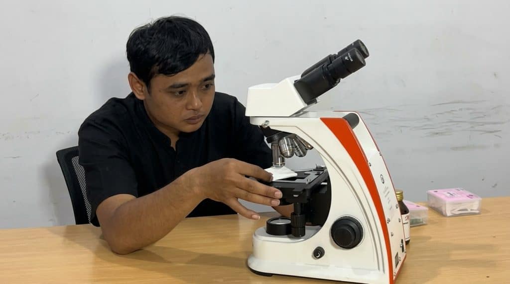
In laboratory activities, visual accuracy is highly important. Sometimes, analysts experience difficulties because the specimen image appears blurry. Therefore, we need to know the standard methods for obtaining clear microscope slide observations. As a result, medical diagnosis and scientific research processes can run smoothly.
Proper Sample and Slide Preparation
The first step begins with the preparation of the specimen itself. Basically, sample thickness heavily affects light penetration. Therefore, ensure that specimen sections or blood smears are made as thin as possible.
Furthermore, the cleanliness of the glass slide(object glass)must not be ignored. Because dust and fingerprint smudges can obstruct the view, always clean the glass using lens tissue. Next, apply the cover slip (cover glass) carefully so that no air bubbles are trapped inside the sample.
Illumination and Condenser Adjustment
The second step is to optimize the device's illumination system. This is because poor lighting will drastically reduce image contrast. First, turn on the illuminator lamp with sufficient intensity.
Subsequently, adjust the position of the condenser and iris diaphragm located under the mechanical stage. If you are using high magnification, raise the condenser closer to the specimen. Then, adjust the diaphragm opening so that the light spreads evenly. In conclusion, an ideal lighting setup will highly support image sharpness.
Focus Adjustment and Lens Calibration
The third step relates to the mechanics of the objective lens. Always start your observation using the objective lens with the lowest magnification (for example, 4x or 10x). Since the field of view is wider, you will find it easier to locate the target position.
After that, use thecoarse adjustmentknob to find the rough image. Next, turn thefine adjustmentknob slowly to achieve a sharp focus. In fact, if you are using a 100x lens, you must apply a drop of immersion oil. This action aims to prevent light refraction that could ruin the final resolution.
Digital Documentation Solutions from PT Miconos
Every laboratory certainly wants perfect evaluation results. Therefore, PT Miconos offers instrument upgrade solutions through the OptiLab microscope line. Additionally, you can equip these microscopes with the Advance Lite or Advance Observer digital camera systems.
Because these devices feature high-resolution sensors, the specimen images can be captured in great detail. Then, those images can be directly displayed on a monitor screen for collaborative evaluation. Does your institution want to improve the quality of its laboratory equipment this year? Immediately visit our official website at www.miconos.co.id or contact a PT Miconos sales representative via WhatsApp right now!